🔥🔥🔥🔥🔥🔥🔥🔥🔥
إضغ بين إيديكم من أقوى الملازم التي صممتها
ملزمة تشريح الجهاز الهيكلي (نظري 3)
💀💀💀💀💀💀💀💀💀💀
تتميز هذهِ الملزمة بعِدة مُميزات :
1- مُترجمة ترجمة تُناسب جميع المستويات
2- تحتوي على 78 رسم توضيحي لكل كلمة موجودة بالملزمة (لكل كلمة !!!!)
#فهم_ماكو_درخ
3- دقة الكتابة والصور عالية جداً جداً جداً
4- هُنالك بعض المعلومات تم توضيحها بشكل تفصيلي جداً (تُعتبر لدى الطالب أو الطالبة بإنها معلومات مُبهمة ومع ذلك تم توضيح هذهِ المعلومات المُبهمة بشكل تفصيلي جداً
5- الملزمة تشرح نفسها ب نفسها بس تكلك تعال اقراني
6- تحتوي الملزمة في اول سلايد على خارطة تتضمن جميع تفرُعات معلومات الجهاز الهيكلي المذكورة في هذهِ الملزمة
واخيراً هذهِ الملزمة حلالٌ عليكم وإتمنى منكم إن تدعولي بالخير والصحة والعافية فقط
كل التوفيق زملائي وزميلاتي ، زميلكم محمد الذهبي 💊💊
🔥🔥🔥🔥🔥🔥🔥🔥🔥
The document provides information about the musculoskeletal system, which is composed of three subsystems: the skeletal system, articular system, and muscular system. It describes the anatomy and functions of the bones, joints, and muscles that make up the axial skeleton (skull, vertebral column, rib cage) and appendicular skeleton (shoulder, pelvis, upper and lower limbs). The skeletal system provides structure, movement, mineral storage, blood cell formation. Typical bones have projections like processes and depressions like foramina that serve attachment and passage functions.
The skeletal system consists of 206 bones and connective tissues that connect them. It performs vital functions like providing a framework to support the body, protecting organs, enabling movement through muscle attachment to bones, producing blood cells, and storing minerals. The skeletal system includes long bones in the limbs, short bones in the hands and feet, flat bones like in the skull, and irregular bones like vertebrae. It is divided into the axial skeleton of the trunk and appendicular skeleton of the extremities. The skull contains cranium and facial bones that protect the brain and house senses.
The document summarizes the skeletal system. It discusses that the skeletal system is composed of bones, cartilage, joints, and ligaments. It then describes the main components of the axial skeleton - the skull, vertebral column, and thoracic cage. The skull is made up of numerous flat and irregular bones that form the cranium and face. The vertebral column consists of 26 vertebrae and intervertebral discs. The thoracic cage is formed by the sternum, ribs, costal cartilages, and thoracic vertebrae.
The skeletal system consists of bones, cartilage, and other connective tissues. It has several functions including support, protection, movement, blood cell formation, and mineral storage. There are different types of bones classified by shape, organization, and histological structure. Long bones have a shaft and two ends, while other bones like those in the skull are flat, irregular, or short. Cartilage is weaker but more flexible than bone and found in joints. The skeletal system includes the axial skeleton of the skull, vertebral column, ribs, and appendicular skeleton of shoulder, pelvis, and limbs.
The document summarizes key aspects of the human skeletal system including its functions of support, movement, protection and blood cell production. It describes the main components of the skeletal system such as bones, joints and ligaments. It provides classifications of bones, joints and muscles. Key facts presented include that the adult human skeleton consists of 206 bones, and examples of the different types of joints like ball-and-socket and hinge joints.
THE SKELETON SYSTEM ANATOMY AND PHYSIOLOGY SLIDESHARE Jitendra Bhargav
This document provides an overview of the skeletal system. It begins with classifying the different types of bones as long, short, flat, or irregular. It then describes the axial and appendicular skeleton in detail, identifying the specific bones that make up each part. Finally, it reviews bone tissue, the process of bone formation, growth and remodeling, and the three types of joints and movements they allow. The skeletal system has important functions of support, protection, movement, mineral storage, and blood cell formation.
There are 206 bones in the human body grouped into the axial skeleton and appendicular skeleton. The axial skeleton includes 80 bones that make up the skull, vertebral column, ribs, and sternum. It forms the central core and foundation of the body. The appendicular skeleton includes 126 bones arranged in the upper and lower limbs, including their attaching girdles, forming the shoulders, arms, legs and allowing for movement.
The document provides information about the human skeletal system including:
1) It describes the two main parts of the vertebrate skeletal system - the axial skeleton which includes the skull, spine and ribs, and the appendicular skeleton which includes the shoulders, arms, hips and legs.
2) It outlines the different bones that make up the human skull including the cranium, facial bones, and auditory ossicles. It also discusses the structure of typical vertebrae and describes the different regions of the vertebral column.
3) It provides details on other bones such as the ribs, sternum, pelvis and the bones that make up the upper and lower limbs. It concludes with an overview of the
The human skeleton is the internal framework of the human body. It is composed of around 270 bones at birth – this total decreases to around 206 bones by adulthood after some bones get fused together. The bone mass in the skeleton makes up about 14% of the total body weight and reaches maximum density around age 21
The skeletal system is composed of bones, cartilage, joints and ligaments. It provides structure and support to the body, protects internal organs, allows for movement via muscle attachment, and stores minerals. The bones are classified as long, short, flat, or irregular. Key bones include the skull, vertebral column, ribs, shoulder girdle, pelvis and bones of the upper and lower limbs. The skeletal system enables vital body functions and works with muscles for movement.
The document provides information about bones and the skeletal system. It discusses the following key points:
- Bones make up the skeletal system and provide structure, protection, movement, mineral storage, blood cell production, and fat storage.
- The skeletal system includes long bones, short bones, flat bones, irregular bones, and sesamoid bones. It is divided into the axial skeleton and appendicular skeleton.
- The axial skeleton includes the skull, vertebral column, ribs, and sternum. It protects organs and allows movement.
- Bones are made of compact bone, spongy bone, periosteum, and marrow. A typical long bone has a diaphysis, epiphyses
This document discusses osteology, the study of the skeleton. It notes that the human skeleton is made up of 206 bones that form the axial and appendicular skeleton. The axial skeleton comprises 80 bones including those of the skull, vertebral column, and thoracic cage. The appendicular skeleton includes 126 bones of the upper and lower limbs along with their attachments. The document classifies bones based on their position, size, shape, gross structure, and development and describes examples for each classification.
The skeletal system provides support and protection for the body’s internal organs and gives the muscles a point of attachment. Humans have an endoskeleton, where our bones lie underneath our skin and muscles. In other animals, such as insects, there is an exoskeleton on the outside of the body.
The skeletal system consists of 206 bones that form the framework of the body. There are two main parts - the axial skeleton along the body's central axis including the skull, vertebral column, and ribcage, and the appendicular skeleton of the limbs. The axial skeleton has 80 bones and protects internal organs. The appendicular skeleton has 126 bones and includes the shoulder and pelvic girdles and upper and lower limbs. Together the skeletal system provides structure, movement, protection, blood cell production, mineral storage, and triglyceride reserves for the body.
At the completion of this unit, learners will be able to: 1. Define skeletal system 2. Discuss the structure, types and functions of bone 3. List the functions of the skeletal system 4. Identify the bones of axial & appendicular skeleton
27
5. Describe the various markings on the surface of bones 6. Describe the bones of: The skull Vertebral column The rib cage or chest Pectoral girdle and upper extremity Pelvic girdle and lower extremity 7. Briefly discuss the difference between male & female pelvis.
Throughout history, the symbol of the skull and crossbones has served as a representation of mortality, likely owing to the fact that following death and decay, bones are the sole remnants. Many individuals perceive bones as inert, desiccated, and fragile. While these attributes accurately portray the bones of a preserved skeleton, the bones within a living human being are profoundly alive. Living bones exhibit strength and flexibility, serving as the primary components of the skeletal system.
The skeletal system comprises 206 bones that support the body and allow for movement. The axial skeleton includes the skull, vertebral column, and thoracic cage, while the appendicular skeleton includes the upper and lower limbs attached to the axial skeleton. The skull protects the brain and is divided into cranial and facial bones. The vertebral column is made up of individual vertebrae that protect the spinal cord and allow for flexibility. Bones are living tissues composed of minerals and connective tissues. The skeletal system provides structure, protects organs, allows body movement, stores minerals, and produces blood cells.
The skeletal system consists of both axial and appendicular parts. The axial skeleton includes the skull, vertebral column, ribs, and sternum. The skull contains both single and paired bones that protect the brain and form cavities for sensory organs. The vertebral column is made up of vertebrae which vary based on region. The appendicular skeleton connects the limbs to the axial skeleton and includes the shoulder girdle, upper limbs, pelvic girdle and lower limbs. Long bones have a shaft and two ends, and different bone types serve various protective and movement functions.
The document discusses the structure and functions of bones in the human skeletal system. It notes that bone is living tissue that is constantly being recreated. Bones provide structure and protection to the body, allow for movement through attachment of muscles and acting as levers, and store minerals and produce blood cells. The five categories of bone are defined based on shape: long bones, short bones, flat bones, irregular bones, and sesamoid bones.
The skeletal system consists of 206 bones that are divided into the axial skeleton (skull, vertebral column, ribs, sternum) and appendicular skeleton (limbs and their attaching girdles). Bones provide structure, protection, movement, mineral storage, blood cell formation, and are living tissues that undergo remodeling. The skeletal system includes various bone cell types and bone is composed of inorganic minerals and organic matrix. Common diseases include osteoporosis, rickets, osteomalacia, and Paget's disease.
QUICK guide to Skeletal System- Dr Gurjant Singh (PT)Dr. Gurjant Singh
So you have read well about human skeletal system and now just want to revise or go through it quickly? Here is the perfect one for you
Dont forget to like and follow for more presentations
Introduction to the Skeletal System.pptxpetrenacolum1
The skeletal system consists of 206 bones that provide structure, protect organs, allow movement, and produce blood cells. There are four types of bones: long bones like the femur; short bones like wrist bones; flat bones like the skull; and irregular bones like vertebrae. The skeletal system works with muscles to support the body, enable movement through leverage, and shield delicate tissues.
The skeletal system has several key functions:
1. It provides structure and support for the body, protects vital organs, and allows for movement.
2. Bones act as levers for muscle attachments and movement.
3. Bones store minerals and act as sites for blood cell formation.
4. There are 206 bones in the adult skeleton divided into long, short, flat, irregular, and sesamoid shapes serving different functions. Bones are classified as either axial or appendicular. Surface markings on bones provide attachment points for muscles and allow for vascular and nerve passageways.
Similar to skeleton System.pdf (skeleton system wow) (20)
A Free 200-Page eBook ~ Brain and Mind Exercise.pptxOH TEIK BIN
(A Free eBook comprising 3 Sets of Presentation of a selection of Puzzles, Brain Teasers and Thinking Problems to exercise both the mind and the Right and Left Brain. To help keep the mind and brain fit and healthy. Good for both the young and old alike.
Answers are given for all the puzzles and problems.)
With Metta,
Bro. Oh Teik Bin 🙏🤓🤔🥰
Post init hook in the odoo 17 ERP ModuleCeline George
In Odoo, hooks are functions that are presented as a string in the __init__ file of a module. They are the functions that can execute before and after the existing code.
How to Download & Install Module From the Odoo App Store in Odoo 17Celine George
Custom modules offer the flexibility to extend Odoo's capabilities, address unique requirements, and optimize workflows to align seamlessly with your organization's processes. By leveraging custom modules, businesses can unlock greater efficiency, productivity, and innovation, empowering them to stay competitive in today's dynamic market landscape. In this tutorial, we'll guide you step by step on how to easily download and install modules from the Odoo App Store.
THE SACRIFICE HOW PRO-PALESTINE PROTESTS STUDENTS ARE SACRIFICING TO CHANGE T...indexPub
The recent surge in pro-Palestine student activism has prompted significant responses from universities, ranging from negotiations and divestment commitments to increased transparency about investments in companies supporting the war on Gaza. This activism has led to the cessation of student encampments but also highlighted the substantial sacrifices made by students, including academic disruptions and personal risks. The primary drivers of these protests are poor university administration, lack of transparency, and inadequate communication between officials and students. This study examines the profound emotional, psychological, and professional impacts on students engaged in pro-Palestine protests, focusing on Generation Z's (Gen-Z) activism dynamics. This paper explores the significant sacrifices made by these students and even the professors supporting the pro-Palestine movement, with a focus on recent global movements. Through an in-depth analysis of printed and electronic media, the study examines the impacts of these sacrifices on the academic and personal lives of those involved. The paper highlights examples from various universities, demonstrating student activism's long-term and short-term effects, including disciplinary actions, social backlash, and career implications. The researchers also explore the broader implications of student sacrifices. The findings reveal that these sacrifices are driven by a profound commitment to justice and human rights, and are influenced by the increasing availability of information, peer interactions, and personal convictions. The study also discusses the broader implications of this activism, comparing it to historical precedents and assessing its potential to influence policy and public opinion. The emotional and psychological toll on student activists is significant, but their sense of purpose and community support mitigates some of these challenges. However, the researchers call for acknowledging the broader Impact of these sacrifices on the future global movement of FreePalestine.
Information and Communication Technology in EducationMJDuyan
(𝐓𝐋𝐄 𝟏𝟎𝟎) (𝐋𝐞𝐬𝐬𝐨𝐧 2)-𝐏𝐫𝐞𝐥𝐢𝐦𝐬
𝐄𝐱𝐩𝐥𝐚𝐢𝐧 𝐭𝐡𝐞 𝐈𝐂𝐓 𝐢𝐧 𝐞𝐝𝐮𝐜𝐚𝐭𝐢𝐨𝐧:
Students will be able to explain the role and impact of Information and Communication Technology (ICT) in education. They will understand how ICT tools, such as computers, the internet, and educational software, enhance learning and teaching processes. By exploring various ICT applications, students will recognize how these technologies facilitate access to information, improve communication, support collaboration, and enable personalized learning experiences.
𝐃𝐢𝐬𝐜𝐮𝐬𝐬 𝐭𝐡𝐞 𝐫𝐞𝐥𝐢𝐚𝐛𝐥𝐞 𝐬𝐨𝐮𝐫𝐜𝐞𝐬 𝐨𝐧 𝐭𝐡𝐞 𝐢𝐧𝐭𝐞𝐫𝐧𝐞𝐭:
-Students will be able to discuss what constitutes reliable sources on the internet. They will learn to identify key characteristics of trustworthy information, such as credibility, accuracy, and authority. By examining different types of online sources, students will develop skills to evaluate the reliability of websites and content, ensuring they can distinguish between reputable information and misinformation.
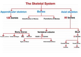
2. The Skeletal System
• Parts of the skeletal system include :
1–Bones (skeleton)
2–Joints
3–Cartilages
4–Ligaments
• Divided into two divisions :
A- Axial skeleton – 80 bones
B- Appendicular skeleton – 126 bones
& X
&
·
/341144
-
SISI
·
12. 3- Flat bones
–Thin and flattened , usually curved
–Thin layers of compact bone around a layer of spongy bone
•Examples: Skull , ribs , sternum
28
-
& -bis
-
-
--
- 26 Je - + /
-2/1204 4:44
13. 4- Irregular bones
–Irregular in shape
–Do not fit into other bone classification categories
Example: Vertebrae and hip
-
-
... &
/
X(2 -5
-
-
-15
-
1 J
,
%
16. 1- Skull
• The cranial and facial bones protect and support special sense organs and
the brain.
• Besides forming the large cranial cavity, the skull also forms several
smaller cavities
– Nasal cavity
– Orbits (eye sockets)
– Para nasal sinuses
– Small cavities which house organs involved in hearing and equilibrium
• Consists of 22 bones
·11 d 160 > // , &
2351
! &
.
111 4
1
j
S
ni 5
.. 97j
- 116141
.
%
+ /
& 3
..j
; 1 + 1 +
= 19
-Si
17.
18. • Bones of the skull are grouped into two categories :
1- Cranial bones : Eight cranial bones form the cranial cavity
a - Frontal bone
b - two parietal bones
c - two temporal bones
d - occipital bone
e - sphenoid bone
f - ethmoid bone
-
......
/19 & .
z
.
11 ...
·
-St
-4
, 15.
-
-
55/
5
./
19. • Frontal Bone :
— Forms the forehead
• Parietal Bones :
— Form the sides and roof of the cranial cavity
• Temporal Bones :
— Form the lateral aspects and floor of the cranium
• Occipital Bone :
— Forms the posterior part and most of the base of the cranium
• Sphenoid Bone :
— Lies at the middle part of the base of the skull
·
.I
-
&.../
2-
1x.11 e
*
- 2
.
%
.
11 &
..
j
-12 /
& Wi ..3
/
51.1 /
&
i
. &/1 , .
/1 g :5 %
.
8
.
/
5541 /
&
. &11 1 / :55 %
.
%
.
1
20. • Ethmoid Bone :
1- A major superior supporting structure of the nasal cavity
2- Contain thin projections called conchae which are lined by mucous membranes
3- location : on the midline in the anterior part of the cranial floor medial to the orbits
4- function : Increased surface area in the nasal cavity helps to humidify inhaled air
-
,9940 4x ..
-
5
. 5
:
=
/36 .-
.
. x
& &
dj ·
&11
. 28 .
30
· 5.
-
.
j
-
156)
21.
22. 2- Facial bones : Fourteen facial bones form the face
a - Two nasal bones
b - two maxillae
c - two zygomatic bones
d - the mandible
e - two lacrimal bones
f - two palatine bones
g - two inferior nasal conchae and vomer
-51 es 14 & 5
6 /
-
11
-
"
1 b.
29. Sutures of the Skull
Sutures : are a type of fibrous joint that are unique to the skull. They are
immovable and fuse completely around the age of 20.
- 1 25
% 1961
[1 %
... 2
.
2
.
1.
=
/
30. • These joints are important in the context of trauma, as they represent points
of potential weakness in the skull. The main sutures in the adult skull are:
1- Coronal suture : fuses the frontal bone with the two parietal bones.
2- Sagittal suture : fuses both parietal bones to each other.
3- Lambdoid suture : fuses the occipital bone to the two parietal bones.
· - 18
.
4 b.
-d 1 <
51
↑ &
*
&
.
.
&
.
32. 2- Vertebral Column
• Also called the spine , backbone , or spinal column
Functions :
1- Protect the spinal cord
2- Support the head
3- Serve as a point of attachment for the ribs, pelvic girdle, and muscles
Sky) -
-8/
-1 di
-
. . . [201p -
33. • The vertebral column is curved to varying degrees in different locations
A- Curves increase the column strength
B- Help maintain balance in the upright position
C- Absorb shocks during walking, and help protect the vertebrae from fracture
- -
-
. = 5
.
3 2
S
-- -
- .. long
s 4b2117151 5
-
. = gas-
34. • Composed of a series of bones called vertebrae (Adult=26)
A- 7 cervical are in the neck region
B- 12 thoracic are posterior to the thoracic cavity
C- 5 lumbar support the lower back
D- 1 sacrum consists of five fused sacral vertebrae
E- 1 coccyx consists of four fused coccygeal vertebrae
-
-
-
-/ = =
(1d
& - &
i S
+ -
..
J
-3) &
- i
-2120
35.
36. Intervertebral Discs
• Location : Found between the bodies of adjacent vertebrae
• Function :
1- Form strong joints
2- Permit various movements of the vertebral column
3- Absorb vertical shock
• Vertebrae typically consist of :
1- A Body (weight bearing)
2- A vertebral arch (surrounds the spinal cord)
3- Several processes (points of attachment for muscles)
Body
- . . / -
(5)
... 9 -
. 95.51i
-5
%
↑
2
-
6) -x14 - )
-
.
/
1 -
=
-5
-
.
37. Types of Discs
1 - Normal Disc
2 - Degenerative Disc
3 - Bulging Disc
4 - Herniated Disc
5 - Thinning Disc
6 - Disc Degeneration with Osteophyte formation
%>81
:4
...
...
·..
S
38.
39.
40. 3- Thorax
• Thoracic cage is formed by the :
1- Sternum
2- Ribs
3- Costal cartilages
4- Thoracic vertebrae
Functions :
1- Enclose and protect the organs in the thoracic and abdominal cavities.
2- Provide support for the bones of the upper limbs.
3- Play a role in breathing.
- 1 1 -
-
-i
&X
T
& ..,
-- -it
T
& s & s y &-1 . . I .. G
S
. >X/ 9 & 1 bX/
-
& Tyga &.....
41.
42. 1- Sternum : *Breastbone*
Location : in the center of the thoracic wall
• Consists of the manubrium, body, xiphoid process
/
· &
s - 113
.
-5- -
·
.. / 2
.
3
.
j)
-
43. 2- Ribs :
• 12 Twelve pairs of ribs give structural support to the sides of the thoracic cavity
• True ribs (7 pairs), False ribs (3 Pairs) and floating ribs(2 pairs)
[04
· 6151 51) %,
·...It &8/1 -
-
/
-
44.
45. 3- Costal cartilages :
• Costal cartilages contribute to the elasticity of the thoracic cage
15
. /
- /
 paypay商城
 乐天二手
乐天二手 日本亚马逊
日本亚马逊 乐天新品
乐天新品 ZOZOTOWN